
LUNGS
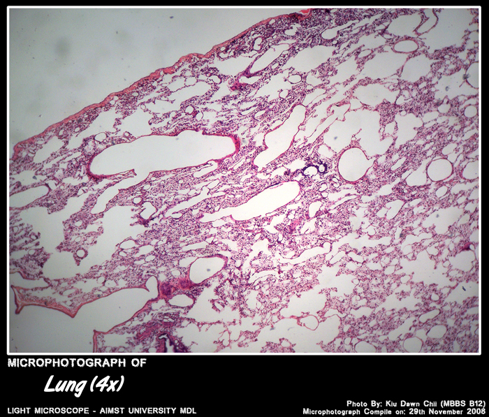
Micro-photograph of Lung tissue under light microscope magnification x4
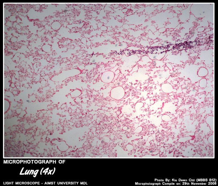
Micro-photograph of Lung tissue under light microscope magnification x4

Micro-photograph of Lung tissue under light microscope magnification x4
Micro-photograph of Lung under light microscope magnification 4x
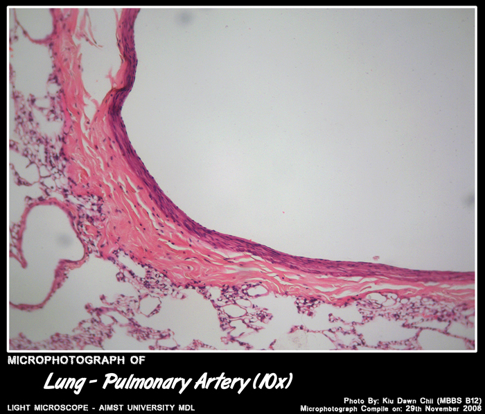
Micro-photograph of Pulmonary artery in lung under light microscope magnification x4
Histology of Bronchi: Primary, secondary, tertiary
- Epithelium – respiratory epithelium – columnar cells have gradual decrease in height, cilia & goblet cells.
- Lamina propria – gradual decrease in thickness & increase in number of elastic fibres.
- Glands – mucous & serous glands .
- Skeletal connective tissue – complete rings in primary, plates of cartilage in smaller bronchi.
- Muscle – several layers of circular smooth muscle.

Micro-photograph of Pulmonary arteries under light microscope magnification 4x
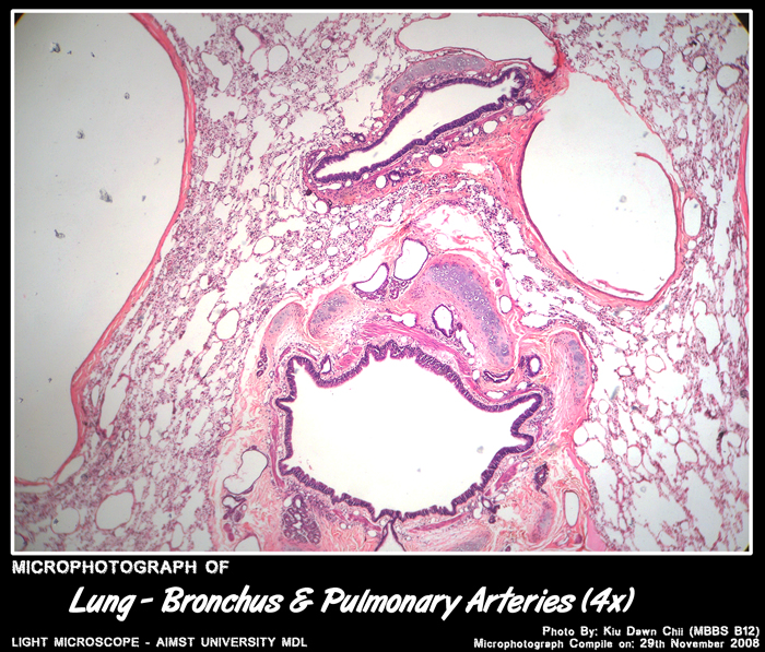
Micro-photograph of Bronchus and pulmonary arteries under light microscope magnification 4x
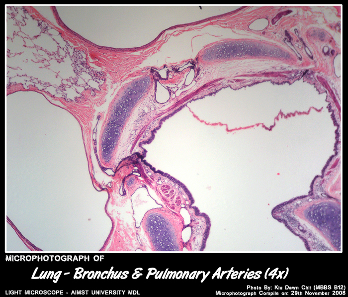
Micro-photograph of Bronchus and pulmonary arteries under light microscope magnification 4x
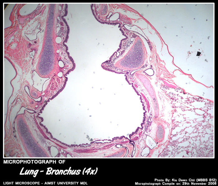
Micro-photograph of Bronchus under light microscope magnification 4x
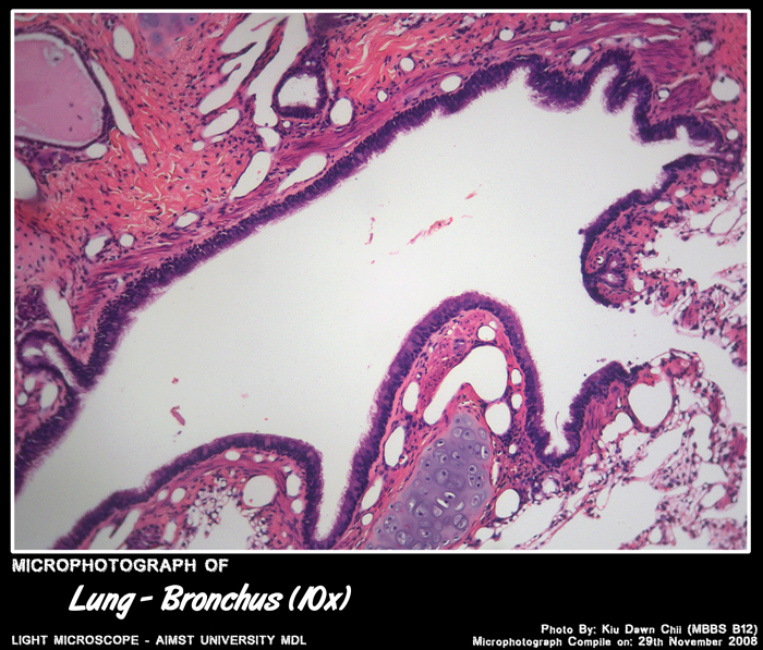
Micro-photograph of Bronchus under light microscope magnification 10x
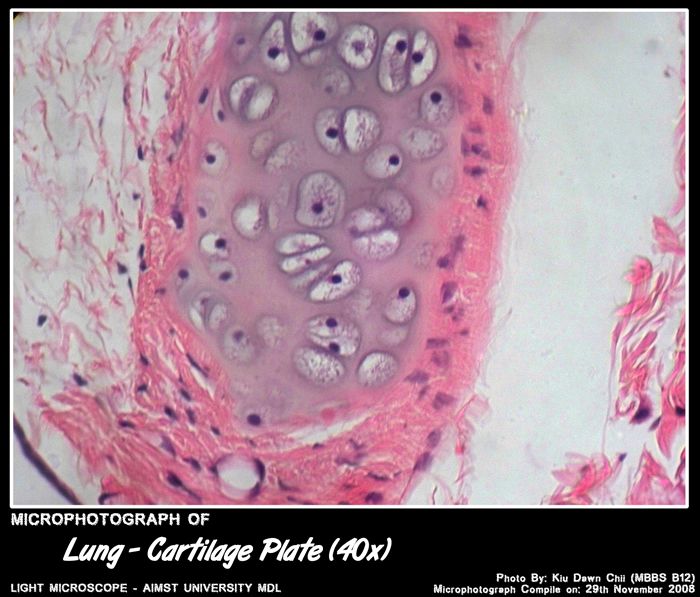
Micro-photograph of Cartilage plate of bronchus under light microscope magnification 40x
Histology of Bronchioles
- Epithelium – Simple columnar with cilia & goblet cells.
- Lamina propria – gradual decrease in thickness & increase in number of elastic fibres.
- Glands – none.
- Skeletal connective tissue – none.
- Smooth muscle – decreasing numbers of smooth muscle cells.

Micro-photograph of Bronchiole under light microscope magnification 10x
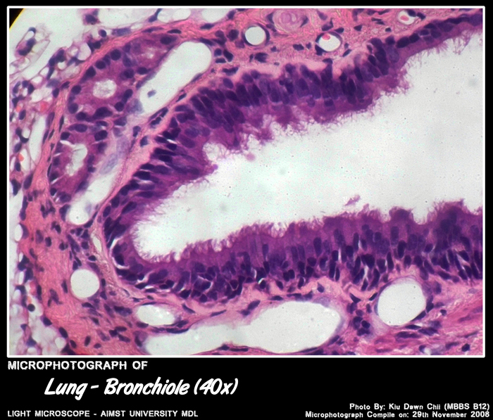
Micro-photograph of Bronchiole under light microscope magnification 40x
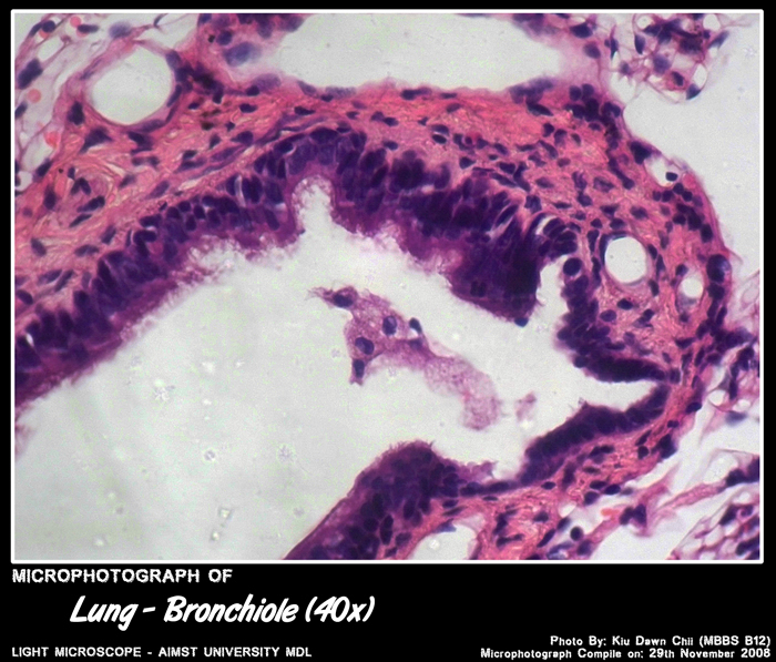
Micro-photograph of Bronchiole under light microscope magnification 40x
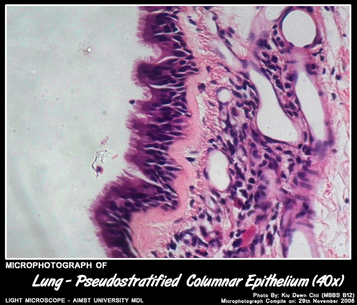
Micro-photograph of Pseudostratified Columnar Epithelium under light microscope magnification 40x
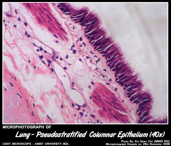
Micro-photograph of Pseudostratified Columnar Epithelium under light microscope magnification 40x
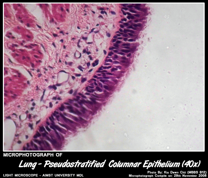
Micro-photograph of Pseudostratified Columnar Epithelium under light microscope magnification 40x
Histology of Terminal bronchioles
- Epithelium – Simple cuboidal cilia free cells named Clara cells , some cells ciliated, goblet cells rare.
- Lamina propria – gradual decrease in thickness & increase in number of elastic fibres.
- Glands – none.
- Skeletal connective tissue – none.
- Smooth muscle – decreasing numbers of smooth muscle cells.
Histology of Respiratory bronchioles
- Epithelium – Low cuboidal, with few cilia; no goblet cells.
- Lamina propria – gradual decrease in thickness & increase in number of elastic fibres.
- Glands – none.
- Skeletal connective tissue – none.
- Smooth muscle – decreasing numbers of smooth muscle cells.
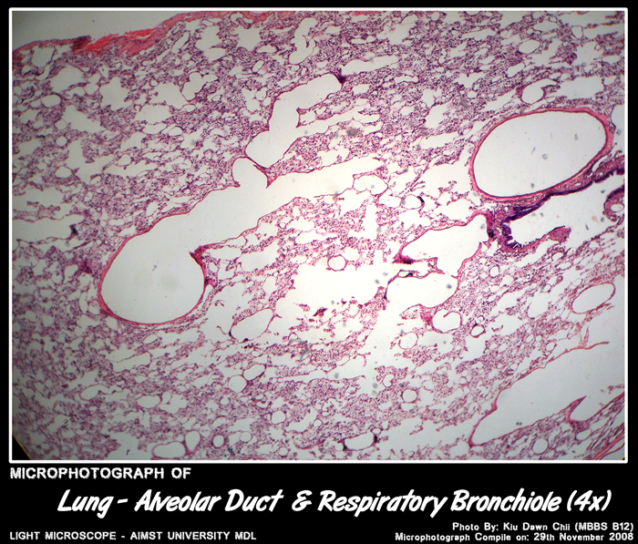
Histology of Alveolar ducts & sacs
- Epithelium – some low cuboidal, no cilia.
- Lamina propria – gradual decrease in thickness & increase in number of elastic fibres.
- Glands – none.
- Skeletal connective tissue – none.
- Smooth muscle – decreasing numbers of smooth muscle cells.
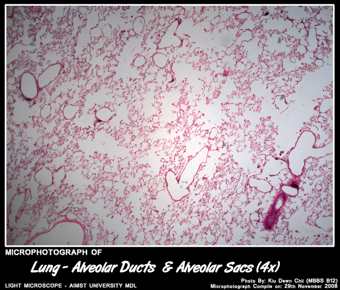
Micro-photograph of Alveolar Ducts and Alveolar Sacs under light microscope magnification 4x

Micro-photograph of Alveolar Ducts and Alveolar Sacs under light microscope magnification 10x
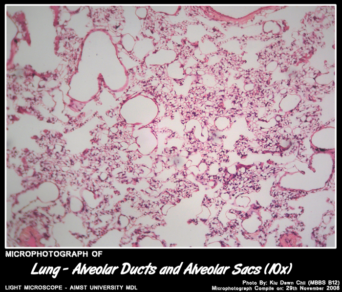
Micro-photograph of Alveolar Ducts and Alveolar Sacs under light microscope magnification 10x
Histology of Alveoli
Epithelium – mostly simple squamous (type 1) – some low cuboidal ( type 2 ) in septa.- Lamina propria very thin with interstitium rich in capillaries & elastic fibres.
- Glands – none.
- Skeletal connective tissue – none.
- Smooth muscle – none.
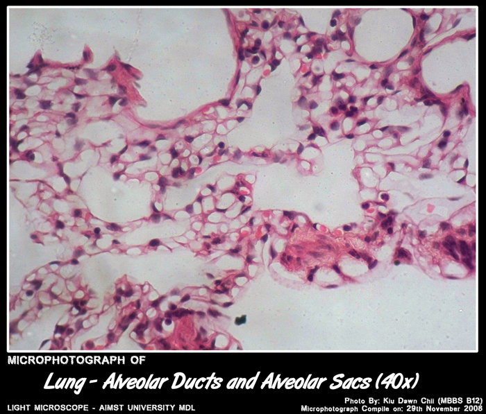
Micro-photograph of Alveolar Ducts and Alveolar Sacs under light microscope magnification 40x
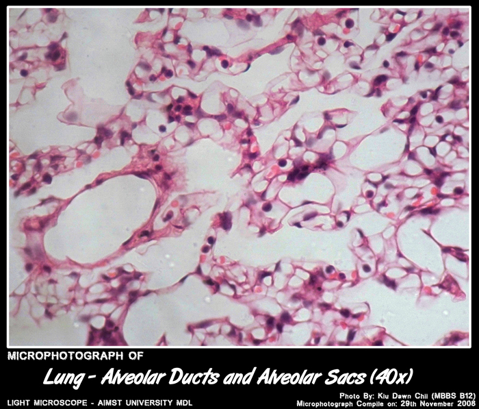
Micro-photograph of Alveolar Ducts and Alveolar Sacs under light microscope magnification 40x
Adapted from: http://myaimst.net/mbbsb12/photo/histo/yr2histo/lung.html
Micro-photograph taken at AIMST University Multi Disciplinary Laboratory during Histology class, using Canon A40 camera over light microscope.